eo_logo
 
Product added to cart

OPTICS ENABLING Life Sciences & Medical Devices

OPTICS ENABLING
Life Sciences &
Medical Devices

Flow Cytometry        Fluorescence Imaging        OCT        COVID-19

Optical Components Enabling Life Science & Medical Device Applications

Optical components from Edmund Optics® are used in countless Life Science-related applications and medical devices such as qPCR instruments, fluorescence microscopes (confocal, multi-photon, super-resolution), flow cytometers (cell sorting), ophthalmoscopes, optical coherence tomography (OCT), and many more. The optics in such devices enable the detection, diagnosis and treatment of conditions such as COVID-19, cancers, macular degeneration, diabetic retinopathy, glaucoma, genetic disorders, hormonal imbalances in the brain, apoptosis in the blood, etc.

Edmund Optics offers all of the optical components you need for building medical devices such as high precision filters, lenses, mirrors, beamsplitters, polarizers, microscope objectives, and more. In addition, Edmund Optics' portfolio comprises a range of light sources and cameras to complete your setup whether you are designing a microscope, an OCT system, or a different type of optical medical device.

  • Nearly 2 million optical components in stock ready to ship for quick system integration and prototyping
  • Custom component design and manufacturing from prototype to full-volume production
  • Key products available from industry leading partners such as SCHOTT, Mitutoyo, Olympus, Nikon, Coherent®, and Hamamatsu
  • ISO9001:2000 and compliance programs designed to meet quality control needs, traceability, and serialization of medical device manufacturers
  • Want an expert technical opinion? Our expert engineers are available for your questions 24/6!

Optics Enabling
Flow Cytometry

Optics Enabling Flow Cytometry

Flow Cytometry is an analytical technique used in a variety of life science applications for counting, inspecting or sorting particles in solution such as single cells. This technique enables the analysis of mixed cell populations - for example from blood, bone marrow, or even from solid tissues such as tumours when these are dissociated into single cells. Flow cytometry is used in a variety of disciplines such as immunology, cancer, virology and molecular biology, as well as infectious disease monitoring. Flow cytometers consist of lenses, optical filters, mirrors, prisms and other optical components to direct the light which are crucial in the successful operation of these systems.

Learn More
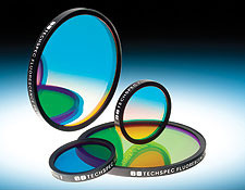
Fluorescence Bandpass Filters

Optical Filters

VIEW ALL
 
FLOW CYTOMETRY
A powerful technology that analyzes physical and chemical characteristics of particles in a fluid suspension. Qualitative and quantitative data is collected as the particles flow through a laser beam and forward and side scattered light is collected.
FLOW CYTOMETRY
 
CELL SORTING
Fluorescence activated cell sorting (FACS) is a specific branch of flow cytometry that actively sorts a heterogeneous collection of cells into various containers a single cell at a time. This is done using general light scattering and fluorescence principles based off of each cell’s characteristics.
CELL SORTING
 
OPTOFLUIDICS
Technology that combines the field of microfluidics with optics. The primary applications include broad covering liquid displays, energy, and optical lenses, but the primary startup company drive is focusing on lab-on-chip devices, biosensors, and molecular imaging systems.
OPTOFLUIDICS HIGH THROUGHPUT SCREENING
A powerful drug-discovery process used heavily in pharmaceuticals. Typically an automated procedure that allows for quicker development of novel drugs with less risk for human error.
HIGH THROUGHPUT SCREENING
 

FLOW CYTOMETRY

A powerful technology that analyzes physical and chemical characteristics of particles in a fluid suspension. Qualitative and quantitative data is collected as the particles flow through a laser beam and forward and side scattered light is collected.

CELL SORTING

Fluorescence activated cell sorting (FACS) is a specific branch of flow cytometry that actively sorts a heterogeneous collection of cells into various containers a single cell at a time. This is done using general light scattering and fluorescence principles based off of each cell’s characteristics.

OPTOFLUIDICS

Technology that combines the field of microfluidics with optics. The primary applications include broad covering liquid displays, energy, and optical lenses, but the primary startup company drive is focusing on lab-on-chip devices, biosensors, and molecular imaging systems.

HIGH THROUGHPUT SCREENING

A powerful drug-discovery process used heavily in pharmaceuticals. Typically an automated procedure that allows for quicker development of novel drugs with less risk for human error.
 
Brain Mapping
Neuroscience technique intended to map and list out the specific quantities or properties of the brain in a spatial representation. In other terms, the anatomy and function of the brain, spine, and central nervous system through imaging techniques.
Brain Mapping
 
Optogenetics
Biological technique that involves the use of light to control cells in living tissue, specifically neurons in most cases that have been genetically modified with photoreceptors that react to different wavebands.
Optogenetics
 
CLARITY
Method of making brain tissue transparent using hydrogels. Accompanied with antibodies or biomarkers, highly detailed pictures of nucleic structure of the brain can be determined and studied.
Clarity
 
GCaMP
A genetically encoded calcium indicator used in brain imaging. GCAMP is similar to the fusion of green fluorescent protein (GFP), calmodulin, and a peptide sequence from myosin.
GCaMP
 
GFP
Green fluorescent protein (GFP) is a specialized protein consisting of a specific group of amino acids that glows green when exposed to UV/Blue light. Extracted from marine life, the most common excitation wavelength is 395nm to 475nm with emission peaks from 509nm to 525nm. GFP is widely used in non-invasive fluorescence imaging systems to detect for tumor growth, apoptosis, and other cellular activity.
GFP
Close up of brain cells

Optics Enabling
Fluorescence Imaging

Optics enabling Fluorescence Imaging

Fluorescence imaging is a powerful, highly sensitive and non-invasive technique used in life sciences to visualise and monitor biological processes in live or fixed cells, tissues, or even complete organisms. Fluorescence is the emission of radiation, visible or invisible, by a substance such as a fluorescent dye (also called fluorophores or chromophores), upon excitation with light or other electromagnetic radiation. There are a wide range of fluorescent dyes and proteins commercially available that can be used to label biological structures with high specificity. Fluorescence-based techniques are varied and include qPCR, DNA sequencing and many microscopy techniques such as confocal, multiphoton and light sheet microscopy. Fluorescence filter sets are particularly important in separating the excitation light from the emitted fluorescence and commonly consist of a combination of an excitation, dichroic and emission filter with transmission profiles optimised to match the spectral characteristics of certain fluorophores.

Learn More
Olympus Water Immersion Objectives

Infinity Corrected Objectives

VIEW ALL

Brain Mapping

Neuroscience technique intended to map and list out the specific quantities or properties of the brain in a spatial representation. In other terms, the anatomy and function of the brain, spine, and central nervous system through imaging techniques.

Optogenetics

Biological technique that involves the use of light to control cells in living tissue, specifically neurons in most cases that have been genetically modified with photoreceptors that react to different wavebands.

Clarity

Method of making brain tissue transparent using hydrogels. Accompanied with antibodies or biomarkers, highly detailed pictures of nucleic structure of the brain can be determined and studied.

GCaMP

A genetically encoded calcium indicator used in brain imaging. GCAMP is similar to the fusion of green fluorescent protein (GFP), calmodulin, and a peptide sequence from myosin.

GFP

Green fluorescent protein (GFP) is a specialized protein consisting of a specific group of amino acids that glows green when exposed to UV/Blue light. Extracted from marine life, the most common excitation wavelength is 395nm to 475nm with emission peaks from 509nm to 525nm. GFP is widely used in non-invasive fluorescence imaging systems to detect for tumor growth, apoptosis, and other cellular activity.

Optics Supporting
optical coherence tomography

Optics Supporting OCT [optical coherence tomography]

Optical coherence tomography (OCT) is a non-invasive, high-resolution optical imaging technology that creates cross-sectional images from interference signals received from an object under investigation and a reference optic. OCT allows improved diagnosis of ophthalmic diseases, such as Age-related Macular Degeneration, AMD (a disease of the retina), which causes blurred vision or diabetic retinopathy, by quantitatively characterising changes in the structure and appearance of retinal tissue. Edmund Optics supplies a wide range of optics ideal for OCT systems, including plate and cube beamsplitters, broadband dielectric mirrors, lenses, illumination sources and whole Lumedica benchtop OCT Imaging Systems.

Learn More
Aspheric Lenses

Optical Lenses

VIEW ALL
 
Ophthalmology
Branch of medicine that deals with the anatomy, study, and diseases of the eye. The eye is one of the leading indicators for diagnosing a number of serious ailments. Due to the ease of access and high level of transmission, the eye has become the “gold standard” for non-invasive medical imaging through various platforms and technologies such as optical coherence tomography (OCT).
Ophthalmology
 
OCT
A powerful medical imaging technique utilizing light to capture high resolution, three-dimensional images from optical scatter in biological tissue. The principles are based off of simple interferometry with near infrared (NIR) light to effectively penetrate the biological medium. A trade-off exists between depth penetration and resolution, but OCT is often coupled with other technologies to ensure accuracy and multi-model images.
OCT
 
BIOMETRICES/ PUPIL RECOGNITION
Automated biometric identification using mathematical algorithms to identify and properly recognize an individual’s iris/pupil. This form of biometric recognition is very reliable, as a human’s eye pattern is completely unique, stable over long periods of time, and can be distinguished and recognized over a great distance.
BIOMETRICS / PUPIL RECOGNITION
 
FUNDUS CAMERA
Low power specialty microscope with a camera acting as a functional ophthalmoscope.
FUNDUS CAMERA

Ophthalmology

Branch of medicine that deals with the anatomy, study, and diseases of the eye. The eye is one of the leading indicators for diagnosing a number of serious ailments. Due to the ease of access and high level of transmission, the eye has become the “gold standard” for non-invasive medical imaging through various platforms and technologies such as optical coherence tomography (OCT).

OCT

A powerful medical imaging technique utilizing light to capture high resolution, three-dimensional images from optical scatter in biological tissue. The principles are based off of simple interferometry with near infrared (NIR) light to effectively penetrate the biological medium. A trade-off exists between depth penetration and resolution, but OCT is often coupled with other technologies to ensure accuracy and multi-model images.

BIOMETRICES/ PUPIL RECOGNITION

Automated biometric identification using mathematical algorithms to identify and properly recognize an individual’s iris/pupil. This form of biometric recognition is very reliable, as a human’s eye pattern is completely unique, stable over long periods of time, and can be distinguished and recognized over a great distance.

FUNDUS CAMERA

Low power specialty microscope with a camera acting as a functional ophthalmoscope.
Optics for
fighting covid-19

The manipulation of light is critical in the fight against COVID-19, which is caused by the novel coronavirus. Frontline healthcare workers and researchers use systems composed of a wide variety of optical components including filters, mirrors, lenses, objectives, and more. These systems include polymerase chain reaction (PCR) devices that replicate DNA samples for testing, antibody detection systems, infrared fever detection devices, UV cleaning robots, and systems for monitoring the blood of patients connected to ventilators.

Learn More
Learn More About Fluorescence Bandpass Filters

Fluorescence Bandpass Filters

VIEW ALL
Learn More About Beamsplitters

Beamsplitters

VIEW ALL
Learn More About Lenses

Lenses

VIEW ALL